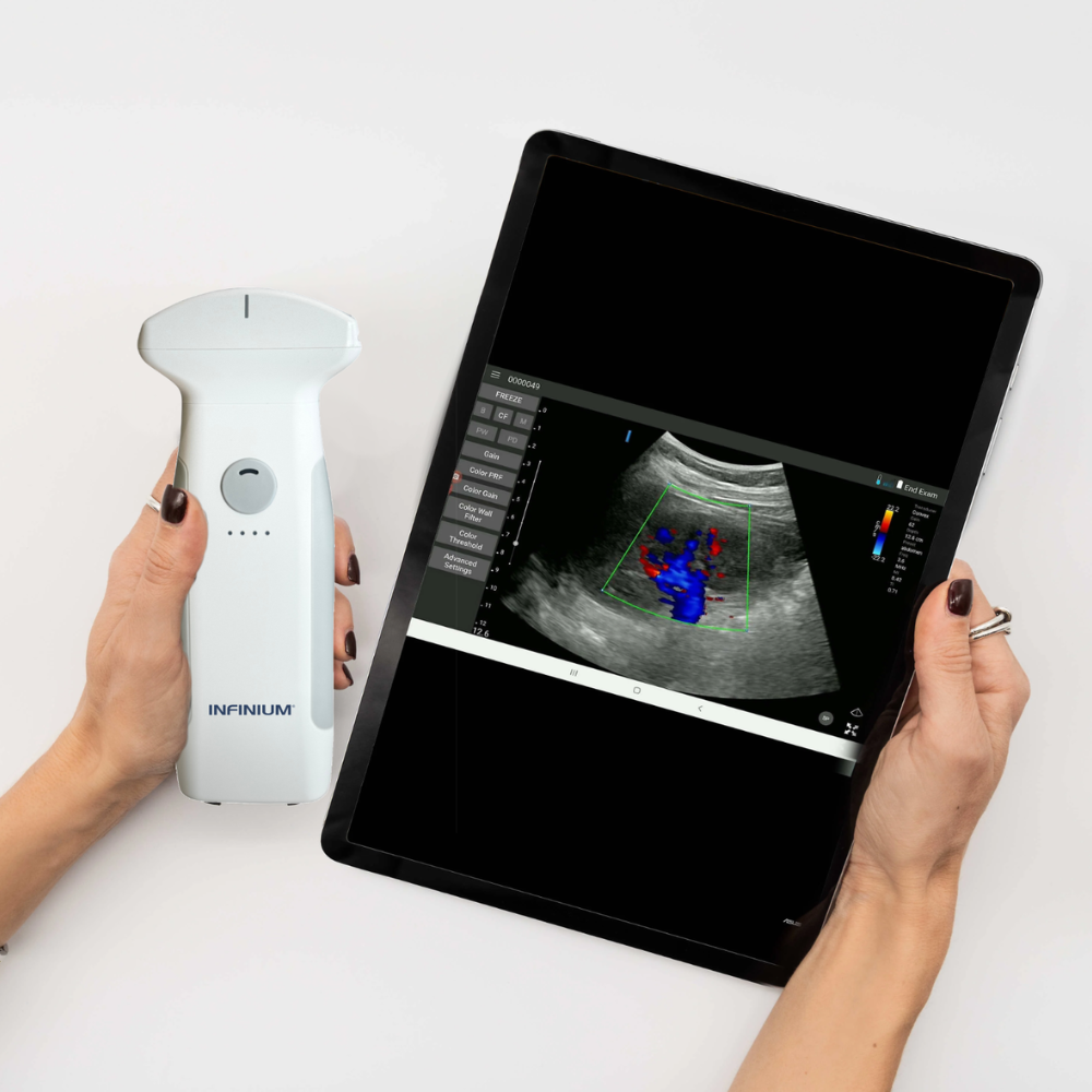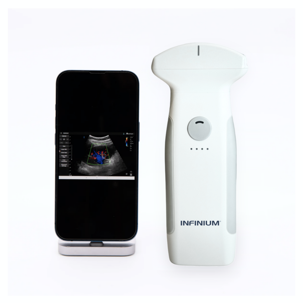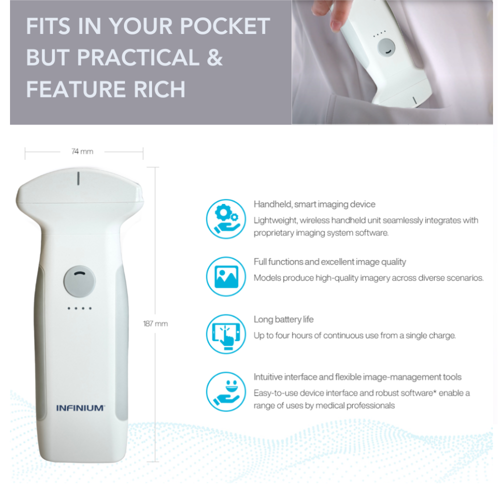

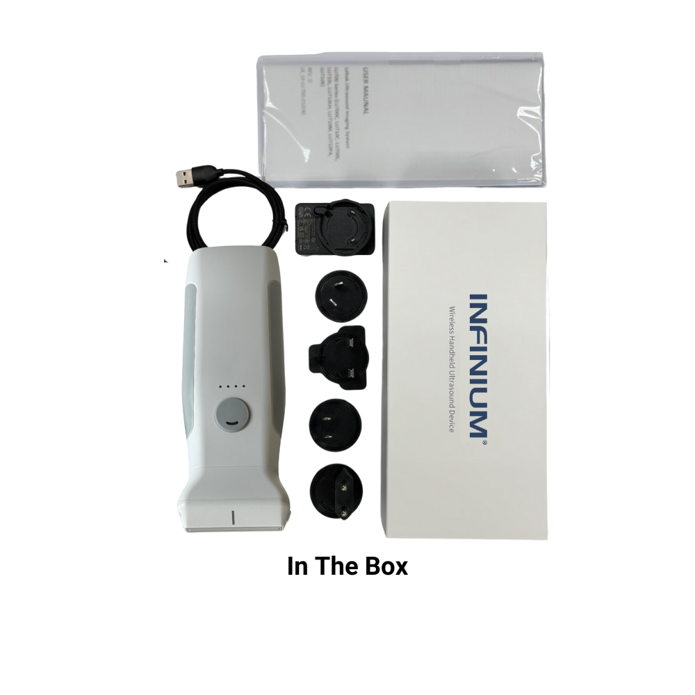
Description
- All
- Abdominal
- Breast
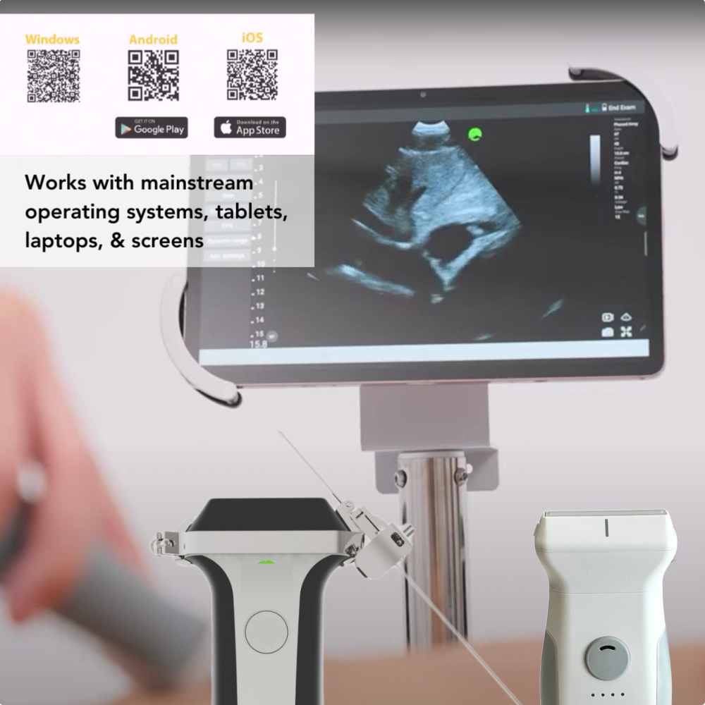
- Heart
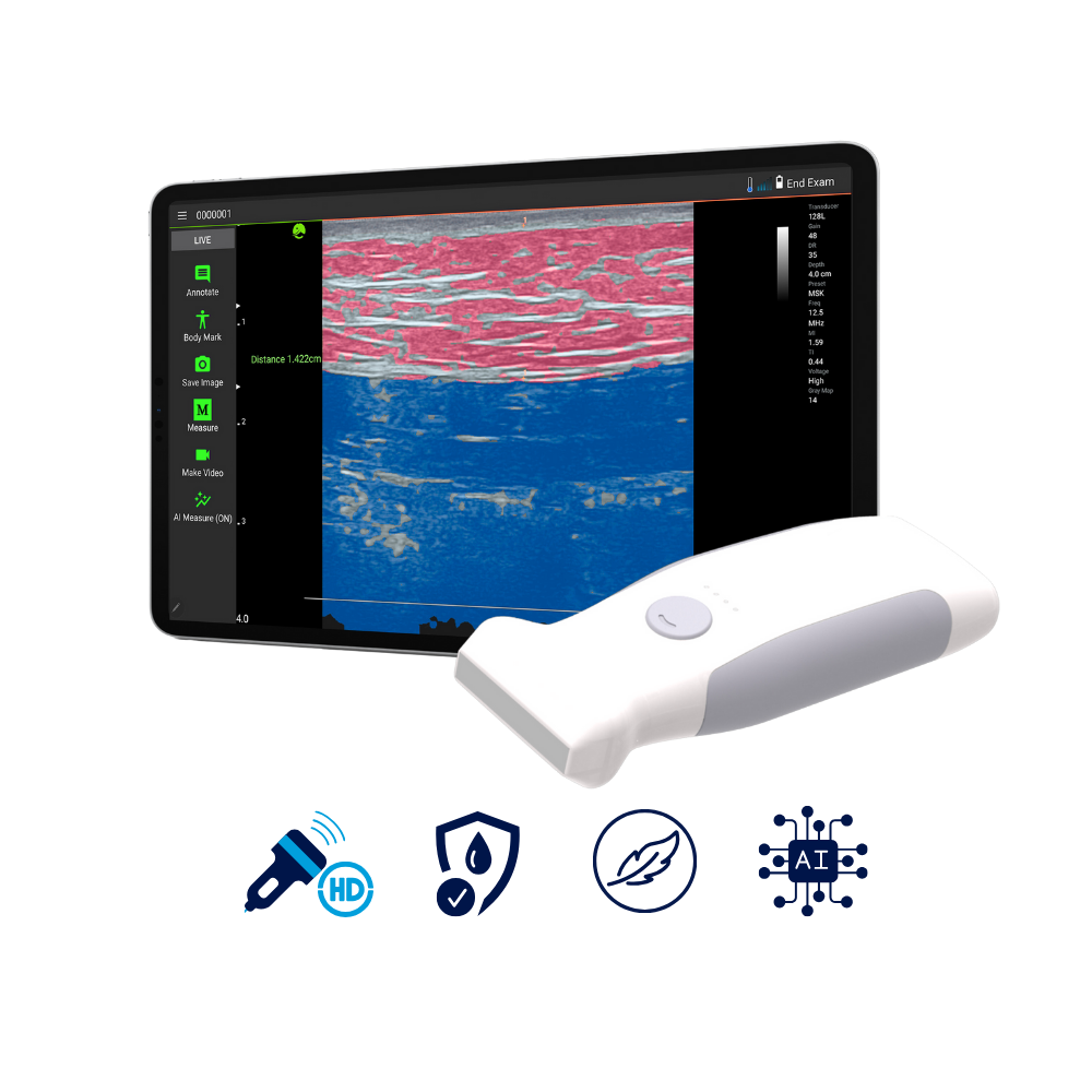
- MSK
- OB
- Vascular
- Other
Kidney
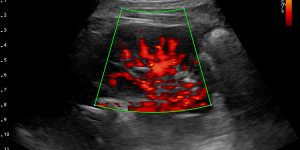
Kidney - Power Doppler
Kidney - Color Doppler
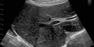
Liver
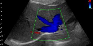
Liver Transverse - Color Doppler
Pancreas
Spleen
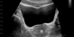
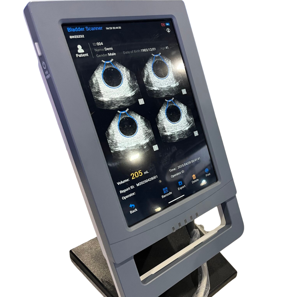
Prostate Gland
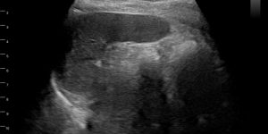
Gall Bladder Stones
Breast Fibroadenoma
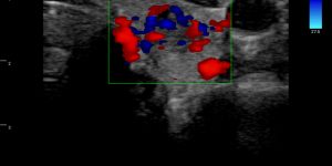
Post Vaccination Axillary Lymph Node Hyperplasia - Color Doppler
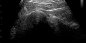
Normal Lung HRUS
Parasternal Short-Axis - M-Mode
Parasternal Long-Axis
Parasternal Short-Axis
Humeral Cartilage
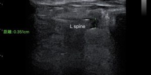
Vertebrae - L Spine
Knee - Spur & Bulging Meniscus
(DJD with femoral spur, bulging of medial meniscus)
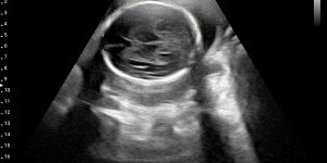
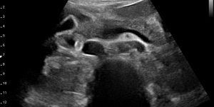
Fetus - 24 Weeks
Fetus - 37 Weeks
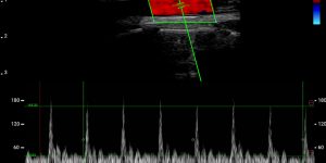
Carotid - Pulse Wave
Finger Peripheral
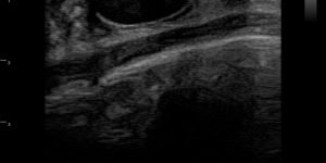
Thyroid
Esophagus
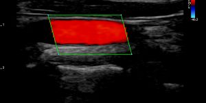
Carotid - Color Doppler
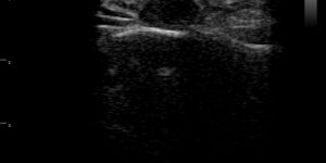
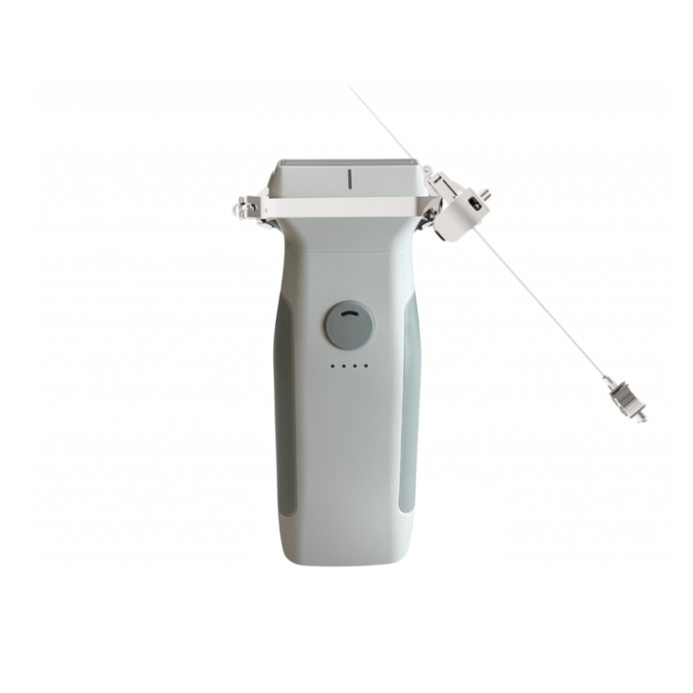
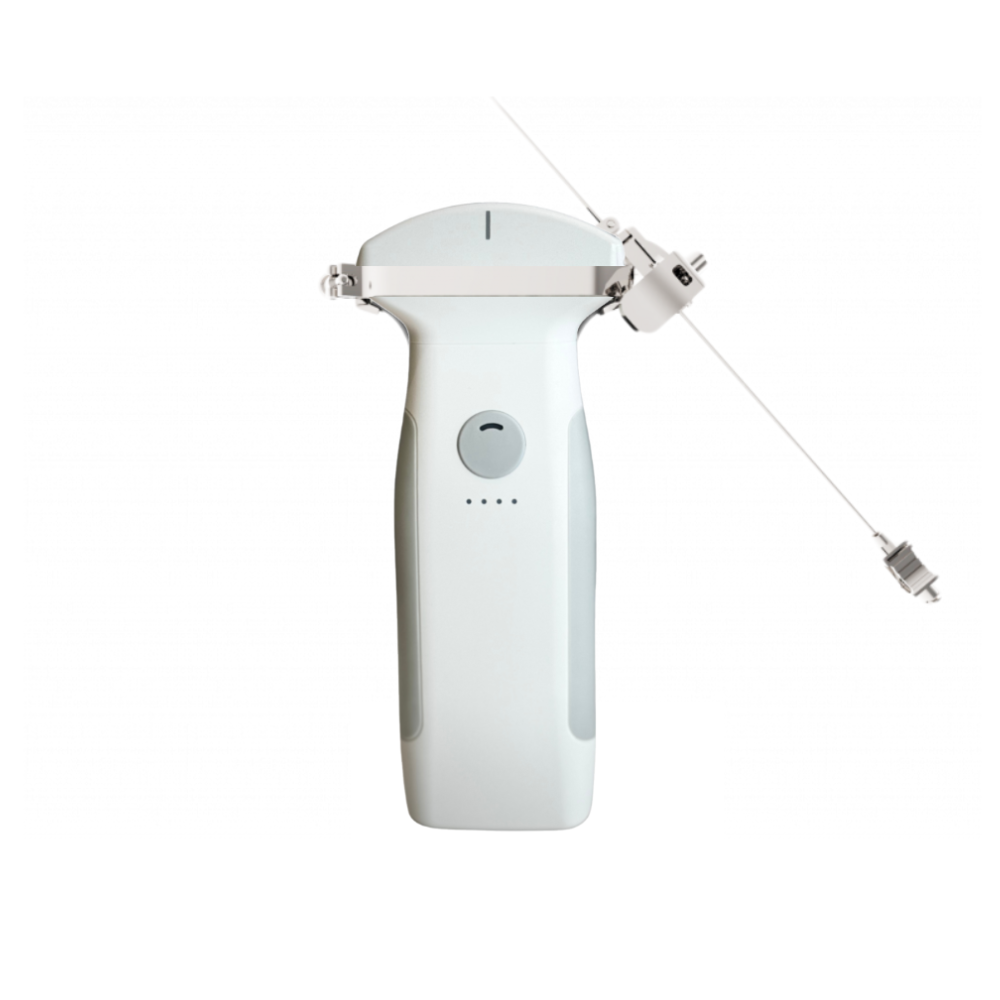
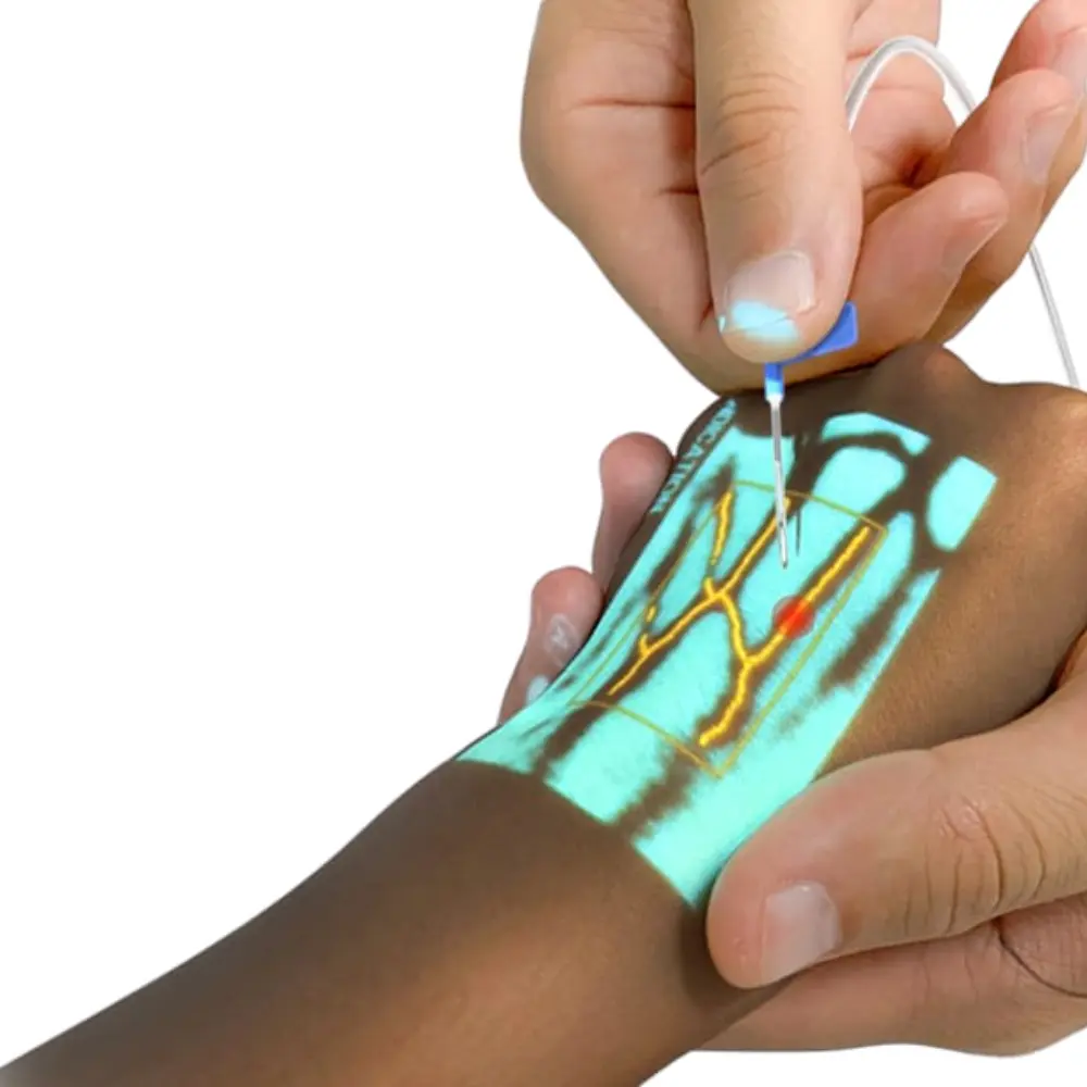
Needle Guided Aspiration